UV harm of Escherichia coli
For survival measurements of E. coli bacteriophage MS2 cells, publish UV irradiation, plaque counting of the cells cultured for twenty-four h on Luria–Bertani (LB) agar plates was carried out as proven in Fig. 1a. The 222-nm irradiation resulted in 1.5-times sooner decay than the 254-nm irradiation. Determine 1b exhibits the 365-nm (0.5 mJ/cm2) photorepair curves of E. coli Ok-12 cells after UV publicity to a dose of 9 mJ/cm2 at 222 and 254 nm. The cells irradiated at 222 nm didn’t recuperate, whereas these at 254 nm did. Publish-photolysis “darkish” restore course of after UV publicity on E. coli Ok-12 at 222 and 254 nm was not noticed throughout 6-h incubation.
(a) Survival charges of E. coli bacterophage MS2 underneath UV irradiation. Log scale base 10. (Black circle) 222 nm, (Black diamond) 254 nm, The susceptibility ratio, okay(222 nm)/okay(254 nm) is 1.5 for the decays. N = 2 and n = 3, (b) (left half)survival charges of E. coli Ok-12 irradiated at 222 and 254 nm, (proper half) photorepair charges by irirradiation at 365 nm. (Black circle) 222-nm irradiation adopted by 365-nm irradiation, (white circle) 222-nm irradiation adopted by preservation underneath darkish situations (management experiment), (black diamond) 254-nm irradiation adopted by 365-nm irradiation, (white diamond) 254-nm irradiation adopted by preservation underneath darkish situations (management experiment). N = 2 and n = 3.
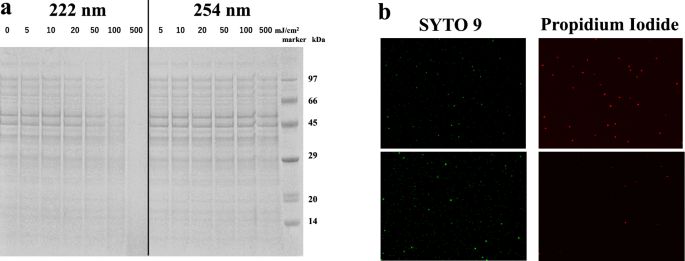
The picture densitometry in Fig. 2a exhibits sodium dodecyl sulfate polyacrylamide gel electrophoresis with DTT added and with out heating to detect signatures for proteins of E. coli Ok-12 (6.9-log CFU/mL). A dose of fifty mJ/cm2 at 222 nm lowered the densitometry depth of the upper molar mass signatures, whereas no vital discount was noticed with a dose as much as 500 mJ/cm2 at 254 nm. The same wavelength dependence on bovine serum albumin was noticed, as proven in Supplementary Fig. S1.
(a) SDS-PAGE of the E. coli K12 protein signature following publicity to growing doses of UV gentle as much as 500 mJ/cm2, as proven in every lane for 222- and 254-nm irradiation. (b) Fluorescent staining of E. coli K12 after UV irradiation. (higher) 222 nm and (decrease) 254 nm with a dose of 200 mJ/cm2.
Cell membrane harm of E. coli Ok-12 was confirmed utilizing the fluorescent staining technique in Fig. 2b with a dose of 200 mJ/cm2. Crimson fluorescence from propidium iodide was dominant within the pattern irradiated at 222 nm, implying that the cells have been useless with compromised cell membranes. The pattern irradiated at 254 nm emitted inexperienced fluorescence from the SYTO9 dye within the cell membranes, implying that the cells had intact cell membranes.
UV harm of a protease, an oligopeptide and amino acids
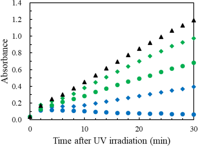
The absorption spectra of aq.options of the frequent fragrant amino acids, tryptophan (Trp), tyrosine (Tyr), phenylalanine (Phe) and histidine (His), are proven in Supplementary Fig. S2. His has absorption at 222 nm however not at 254 nm. Trp and Tyr have robust absorption at each 222 and 254 nm, whereas Phe has a weak absorption. A protease, chymotripsin, catalyses the peptide bond hydrolysis of Bz-Tyr-pNA (BTPNA in 50percentDMSO / 50percentwater) in its S1 binding pocket of His 57, Ser 195 and Asp 102. To evaluate UV inhibition of the catalytic exercise, aq. HCl options of α-chymotripsin have been irradiated at 222 and 254 nm (0.5 mW/cm2) with doses of 100 and 500 mJ/cm2. After UV irradiation to α-chymotripsin, a combination of the α-chymotripsin answer (75 μg/mL) and a BTNPA answer have been monitored at a probe wavelength of 405 nm in a Tris∙HCl buffer answer, probing the hydration product, p-nitroaniline, as proven in Fig. 3. The upper dose and shorter UV wavelength resulted in much less manufacturing of p-nitroaniline. The ratios of the slopes, r = (UV-irradiated)/(management), correspond to the residual catalytic actions. For a UV dose of 100 mJ/cm2, which is obtained from the info between t = 10–20 min in Fig. 3, r(222 nm) is 0.30, whereas r(254 nm) is 0.81 suggesting that the exercise was 3.5 (= 70/19) occasions lowered by 222-nm irradiation than by 254-nm irradiation. The UV dose results on the absorption spectra of the α-chymotripsin options have been measured for does of 0–500 mJ/c2, as proven in Supplementary Fig. S3. The absorption spectra have a peak round 280 nm on account of fragrant amino acid residues (Trp, Tyr and Phe). These chromophores take in 254-nm photons to dysfunction the construction and reduce the catalytic capacity with none considerable change in its absorption spectrum. The spectral change after the 222-nm irradiation was proved at 225 nm (lower of absorbance) and 250 nm (improve). The decreases within the catalytic capacity and absorption spectral depth suggest the photodegradation of the His facet chain within the binding pocket since (a) His not solely stabilises creating fees, but in addition supplies a path for proton switch, with out which catalytic reactions would have issue in continuing, and (b) His is a powerful chromophore at 222 nm.
Manufacturing of p-nitroaniline by monitoring absorption depth at 405 nm after UV irradiation to aq.options of chymotrypsin. UV doses in mJ/cm2 are (Blue diamond) 100 and (Blue circle) 500 at 222 nm, (inexperienced diamond) 100 and (inexperienced circle) 500 at 254 nm, (Black circle) 0. The preliminary hump is brought on by gentle scattering. n = 1.
Regarding the small discount within the catalytic exercise, r, after 254-nm irradiation, His is related to this as a result of its weak photoabsorption. Though the catalytic response was inhibited, Supplementary Fig. S3 exhibits solely a slight change within the UV absorption spectra after 254-nm irradiation. It is because the photoproducts might need a UV spectrum that resembles to the unique α-chymotripsin one. McLaren and Luse19 reported that, of their 254-nm irradiation to chymotrypsin by chemical evaluation, about one Trp residue per chymotrypsin molecule was destroyed and none of disulphide linkages have been damaged. No Phe, Tyr, or His residues have been modified.
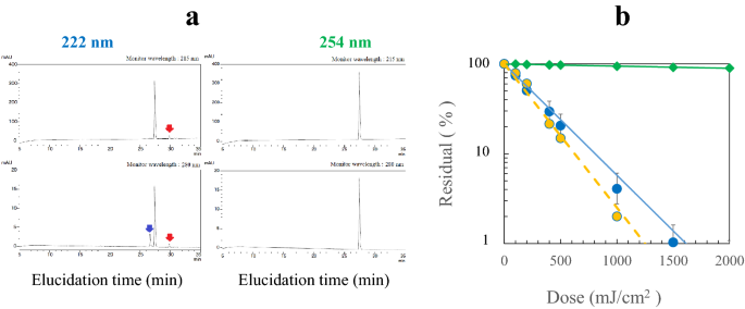
To evaluate the function of the residues additional, an oligopeptide, angiotensin II (Asp-Arg-Val-Tyr-Ile-His-Professional-Phe) in aq.answer (50 μM) was UV irradiated at 222 and 254 nm. The HPLC evaluation for a dose of 100 mJ/cm2 is proven in Fig. 4a. For the 222-nm irradiation, the facet peaks of the photoproduced peptides have been noticed at two monitor wavelengths of 215 and 280 nm, whereas no facet peak for the 254-nm irradiation. The crimson arrows point out the photoproduct assigned to a peptide containing Tyr since they appeared by monitoring at each 215 and 280 nm. The blue one to a peptide containing the merchandise from the photodegradation of His because it seems strongly at 280 nm. Determine 4b exhibits that irradiation at 222 nm induced robust discount, whereas it was very weak at 254 nm. Dose-dependent variations within the HPLC peak intensities are proven in Supplementary Fig. S4. For the dose of 0–1.0 J/cm2, the product depth elevated and the angiotensin II depth decreased. Determine 4b additionally exhibits the deaeration impact of aq.options with N2 fuel effervescent underneath sonication. Deaeration solely barely modified the discount charge.
(a) HPLC elucidation profiles after UV irradiation of angiotensin II with a dose of 100 mJ/cm2 at 222 and at 254 nm. The monitoring wavelengths are (higher) 215 nm, and (decrease) 280 nm. The crimson arrows point out the photoproduct assingned to a peptide containing tyrosine, and the blue one to a peptide containing the product from photodegradation of histidine. (b) discount of angiotensin II by UV irradiation. (Blue circle) 222 nm and (inexperienced diamond) 254 nm with aq. options aerated, (yellow circle) 222 nm with aq. options deaerated. n = 3.
Determine 4b exhibits the wavelength dependence of the discount of angiotensin II with robust discount by irradiation at 222 nm and virtually no discount 254 nm. To evaluate the photodegradation function of the amino acid residues, free Tyr, Trp, Phe and His, aq. options (50 μM) have been irradiated at 222 and 254 nm for doses of 0– 2 J/cm2 in Supplementary Fig. S5. The residual percentages at a dose of 1.0 J/cm2 have been as follows: at 222 nm His (< 1%): Trp (16%): Tyr (38%): Phe (82%), and at 254 nm His (100%):Trp (71%): Tyr (88%): Phe (95%). Tyr is appreciably lowered at 254 nm and ca. eightfold much less lowered than His at 222 nm. Thus, the photoreduction susceptibility to His matches the wavelength dependence of the noticed discount charges of angiotensin II in Fig. 4b, whereas to not Tyr, since free Tyr has considerable absorption at 245 nm and His has no absorption. The deaeration impact of the angiotensin II answer is small in Fig. 4b. Within the oxidation means of free fragrant amino acids, it has been kown that His and Trp react at considerable charges with singlet oxygen20. As proven in Supplementary Fig. S5, the deaeration twofold decreased the UV discount charge for Trp throughout 222-nm irradiation, however not for His. Thus, His residue is essentially the most believable for the UV discount of angiotensin II. The deaeration of aq. answer didn’t afford safety in opposition to His degradation at 222 nm, implying that the first photochemistry at 222 nm is direct photodegradation and unbiased of dissolved O2.
UV harm of plasmid DNA at 222 and 254 nm and photorepair capacity at 365 nm
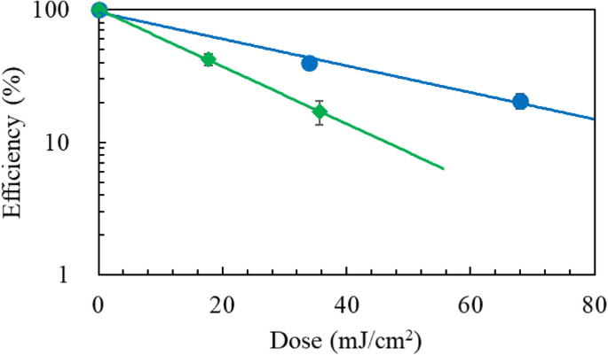
Following UV excitation on DNA, adjoining pyrimidines might type lesions, CPD or (6-4)PP. The CPD lesion is photorepaired by a photolyase and a coenzyme (flavin adenine dinucleotide, FAD) by long-wavelength gentle, whereas the (6-4)PP lesion is just not. We examined UV harm of plasmid DNA (1, 5, 10 pg) in Tris∙EDTA buffer options at 222 and 254 nm, which have been reworked into E. coli HB101 competent cells after UV irradiation. Determine 5 exhibits the transformation effectivity curves, for which the uncooked information of colony counting are listed in Supplementary Desk S1. The doses at 21 and 40% transformation have been 68 and 34 mJ/cm2 for 222 nm, whereas for 254 nm the doses at 17 and 42% transformation have been 36 and 18 mJ/cm2 The photodamage susceptibility of plasmid DNA is twofold decrease at 222 nm than at 254 nm due to the weaker absorbance of DNA at 222 nm14.
To evaluate the photorepair capacity of the broken DNA, two units of plasmid DNA samples that have been lowered to twenty% residual by UV irradiation have been reworked into E. coli in ampicillin LB plates. After 60 min with and with out photorepair irradiation at 365 nm (0.36 mW/cm2), the plates have been positioned for cultivation, after which, colony counting was carried out. The common colony numbers for the 222-nm irradiation have been N222 (with 365 nm) = 61 and N222 (with out 365 nm) = 40. For the 254-nm irradiation, N254 (with 365 nm) = 61 and N254 (with out 365 nm) = 31. The photorepair increment ratio at 222 nm is R222 = N222 (with 365 nm)/N222(with out 365 nm)—1 = 61/40–1 = 0.53, whereas at 254 nm, R254 = 61/31–1 = 0.97. These outcomes suggest that (a) manufacturing of CPD in plasmid DNA irradiated at 222 nm is lower than at 254 nm and/or (b) CPD photorepair by the photolyase is much less energetic at 222 nm than at 254 nm.
Photodamage of a cofactor FAD within the CPD photorepair enzymatic course of
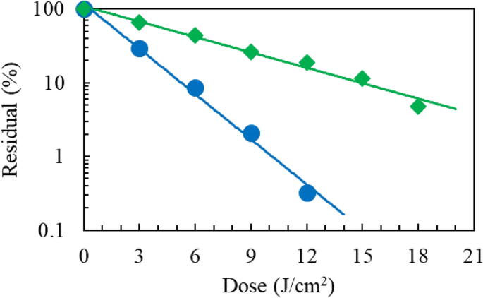
The photolyase harbours an FAD coenzyme to reverse CPD to the adjoining pyrimidines. As proven in Fig. 6, UV irradiation of FAD (25 μg/mL) in aq. answer at 222 nm as much as a dose of 18 J/cm2 induced harm three-fold extra effectively than at 254 nm, implying that CPD photorepairs by the photolyase grew to become much less energetic by irradiation at 222 nm than at 254 nm. HPLC elucidation profiles are proven in Supplementary Fig. S6.
UV harm of nucleosides
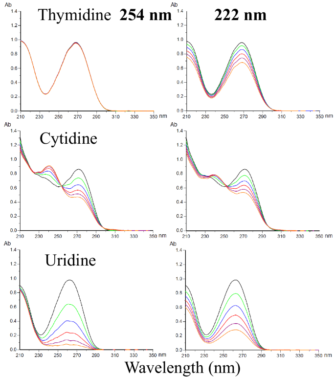
DNA consists of a sugar-phosphate spine to which the 4 nucleobases, adenine (A), guanine (G), cytosine (C), thymine (T), are hooked up by glycosidic bonds, whereas T is changed by uracil (U) in RNA. The absorption spectra of nucleosides have a peak within the vary 250–270 nm, and the spine absorbs at round 210 nm. The photodamage of those nucleosides in aq. answer (50 μM) was investigated by measurement of absorption spectral modifications. As proven in Fig. 7 with doses as much as 5.0 J/cm2, the pyrimidine bases, C and U, have been comparably photodamaged by 222-nm and 254-nm irradiation. The pyrimidine base, T, was photodamaged at 222 nm however not at 254 nm. Thus, T is most believable to the UV harm of plasmid DNA at 222 nm in Fig. 5. Equally, no considerable modifications within the absorption spectrum of the purine nucleobases, adenosine and guanosine, have been noticed upon UV irradiation, as proven in Supplementary Fig. S7. For the reason that mechanism of photo-damage is essentially completely different between nucleoside monomer and plasmid on account of their surroundings distinction, further experiments are wanted to verify these assumptions.
UV harm of RNA UpU
RNA UpU is a mannequin nucleotide block for RNA. UpU (50 μg/mL) in aq.answer was UV irradiated to evaluate the wavelength sensitivities to break of UpU and formation of (6-4)PP and CPD. By HPLC evaluation with monitoring at 258 nm, the discount of UpU by irradiation at 222 and 254 nm as much as a dose of 4.5 J/cm2 was measured, as proven in Fig. 8a. The degradation charge at 222 nm was 1.9 ± 0.1 occasions decrease than that at 254 nm. Determine 8b exhibits that the manufacturing of (6-4)PP from UpU is 1.5-times bigger by irradiation at 222 nm than at 254 nm, implying that the manufacturing of (6-4)PP at 222 nm is 2.9-time (1.9 × 1.5) extra environment friendly than at 254 nm. Supplementary Figs. S8 and S9 present the spectral modifications of UpU by UV irradiation and HPLC elucidation profiles of the merchandise, respectively. The HPLC analyses present {that a} weak CPD sign was noticed for 222-nm irradiation, whereas a powerful one for 254-nm irradiation. Primarily based on these outcomes, the photorepair yield of RNA broken by irradiation at 222 nm is predicted to be low in a germicidal course of.
UV photochemistry of RNA UpU. (Blue circle) 222 nm and (inexperienced diamond) 254 nm (a) discount of UpU as a perform of dose. n = 1, (b) manufacturing of (6-4)PP from UpU as a perform of dose. n = 1, (c) restoration of photohydrated UpU as a result of self-reversion response at room temperature underneath darkish situations after UV irradiation. SD = 2% of every sign depth. n = 3.
UV irradiation of dTpdT produced solely (6-4)PP at 222 nm, whereas each (6-4)PP and CPD at 254 nm, as proven within the HPLC elucidation profiles of Supplementary Fig. S10.
Manufacturing of hydrate from UpU and its “darkish” reversion
Along with strange photodamage processes by UV irradiation, a water molecule provides throughout the pre-existing double bond in a nucleophilic hydrolysis response and is equilibrated with the non-hydrated UpU18. The reversible response of hydrated UpU was examined with these samples subsequently preserved for as much as 120 h at room temperature underneath darkish situations. The self-reversion course of from hydrated UpU to non-hydrated one was monitored at 215-nm absorption of UpU in HPLC evaluation. The elucidation profiles after 254-nm irradiation are proven in Supplementary Fig. S11. Determine 8c exhibits the self-recovery curves of UpU broken by UV irradiation. UpU was broken to a 4% residual (96% harm) by irradiation at 254 nm at a dose of 4.5 J/cm2. After preservation for 120 h, UpU recovered to a 68% residual. Thus, the restoration quantity was 64%. At 222 nm with a dose of seven.2 J/cm2 UpU was broken to a 11% residual (89% harm), which recovered to a 24% residual. Thus, the restoration quantity was 13%.
Notice that the formation of thymine hydrates by 254-nm irradiation has a low quantum yield on account of steric hindrance by the 5-CH3 substituent in thymine. As proven within the HPLC elucidation profiles in Supplementary Fig. S10, UV irradiation of dTpdT at 254 nm produced CPD and (6-4)PP, whereas solely (6-4)PP at 222 nm.
Desk 1 summarizes the comparability of damages and merchandise brought on by 222-nm and 254-nm irradiation on cell proteins, chymotripsin, angiotensin II, plasmid DNA, FAD photorepair coenzyme and uracil/thymine dimer blocks.


