Abstract: Neurons within the midbrain obtain sturdy, particular synaptic enter from retinal ganglion cells, however solely from a small variety of the sensory neurons.
Supply: Charite
For the primary time, neuroscientists from Charité – Universitätsmedizin Berlin and the Max Planck Institute for Organic Intelligence (at the moment within the means of being established) have revealed the exact connections between sensory neurons contained in the retina and the superior colliculus, a construction within the midbrain.
Neuropixels probes are a comparatively latest growth, representing the following technology of electrodes. Densely filled with recording factors, Neuropixels probes are used to file the exercise of nerve cells, and have facilitated these latest insights into neuronal circuits.
Writing in Nature Communications, the researchers describe a basic precept which is frequent to the visible techniques of mammals and birds.
Two mind constructions are essential to the processing of visible stimuli: the visible cortex within the major cerebral cortex and the superior colliculus, a construction within the midbrain. Imaginative and prescient and the processing of visible info contain extremely advanced processes.
In simplified phrases, the visible cortex is answerable for common visible notion, whereas the constructions of the evolutionarily older midbrain are answerable for visually guided reflexive behaviors.
The mechanisms and rules concerned in visible processing inside the visible cortex are well-known. Work performed by a group of researchers led by Dr. Jens Kremkow has contributed to our information on this area and, in 2017, culminated within the institution of an Emmy Noether Junior Analysis Group at Charité’s Neuroscience Analysis Middle (NWFZ).
The first intention of the Analysis Group, which is funded by the Germany Analysis Basis (DFG), is to additional enhance our understanding of nerve cells concerned within the visible system. Many unanswered questions stay, together with particulars of the best way through which visible info is processed within the midbrain’s superior colliculi.
Retinal ganglion cells, sensory cells discovered inside the attention’s retina, reply to exterior visible stimuli and ship the data obtained to the mind. Direct signaling pathways be certain that visible info obtained by the retinal nerve cells additionally reaches the midbrain.
“What had remained largely unknown till now’s the best way through which nerve cells within the retina and nerve cells within the midbrain are linked on a purposeful degree. The dearth of data concerning the best way through which neurons within the superior colliculi course of synaptic inputs was equally pronounced,” says examine lead Dr. Kremkow.
“This info is essential to understanding the mechanisms concerned in midbrain processing.”
Till now, it had been not possible to measure the exercise of synaptically related retinal and midbrain neurons in dwelling organisms. For his or her most up-to-date analysis, the analysis group developed a technique which was based mostly on measurements obtained with progressive, high-density electrodes often known as Neuropixels probes.
Exactly talking, Neuropixels probes are tiny, linear electrode arrays that includes roughly one thousand recording websites alongside a slender shank. Comprising 384 electrodes for the simultaneous recording of electrical exercise of neurons within the mind, these gadgets have turn into game-changers inside the area of neuroscience.
Researchers working at Charité and the Max Planck Institute for Organic Intelligence have now used this new know-how to find out the related midbrain constructions in mice (superior colliculi) and birds (optic tectum).
Each mind constructions have a typical evolutionary origin and play an vital function within the visible processing of retinal enter alerts in each teams of animals.
Their work led the researchers to a stunning discovery: “Normally, one of these electrophysiological recording measures electrical alerts from motion potentials which originate within the soma, the neuron’s cell physique,” explains Dr. Kremkow.
“In our recordings, nonetheless, we observed alerts whose look differed from that of regular motion potentials. We went on to analyze the reason for this phenomenon, and located that enter alerts within the midbrain had been attributable to motion potentials propagated inside the ‘axonal arbors’ (branches) of the retinal ganglion cells. Our findings counsel that the brand new electron array know-how can be utilized to file {the electrical} alerts emanating from axons, the nerve cell projections which transmit neuronal alerts. It is a brand-new discovering.”
In a worldwide first, Dr. Kremkow’s group was in a position to concurrently seize the exercise of nerve cells within the retina and their synaptically related goal neurons within the midbrain.
Till now, the purposeful wiring between the attention and midbrain had remained an unknown amount. The researchers had been in a position to present on the single-cell degree that the spatial group of the inputs from retinal ganglion cells within the midbrain constitutes a really exact illustration of the unique retinal enter.

“The constructions of the midbrain successfully present an virtually one-to-one copy of the retinal construction,” says Dr. Kremkow.
He continues: “One other new discovering for us was that the neurons within the midbrain obtain a really sturdy and particular synaptic enter from retinal ganglion cells, however solely from a small variety of these sensory neurons. These neural pathways allow a really structured and purposeful connection between the attention’s retina and the corresponding areas of the midbrain.”
Amongst different issues, this new perception will improve our understanding of the phenomenon often known as blindsight, which may be noticed in people who’ve sustained injury to the visible cortex attributable to trauma or tumor.
Incapable of acutely aware notion, these people retain a residual potential to course of visible info, which ends up in an intuitive notion of stimuli, contours, motion and even colours that seems to be linked to the midbrain.
To check whether or not the rules initially noticed within the mouse mannequin might additionally apply to different vertebrates – and therefore whether or not they may very well be extra common in nature – Dr. Kremkow and his group labored alongside a group from the Max Planck Institute for Organic Intelligence, the place a Lise Meitner Analysis Group led by Dr. Daniele Vallentin focuses on neuronal circuits answerable for the coordination of exact actions in birds.
“Utilizing the identical sorts of measurements, we had been in a position to present that, in zebra finches, the spatial group of the nerve tracts connecting the retina and midbrain observe the same precept,” says Dr. Vallentin.
She provides: “This discovering was stunning, provided that birds have considerably increased visible acuity and the evolutionary distance between birds and mammals is appreciable.”
The researchers’ observations counsel that the retinal ganglion cells in each the optical tectum and the superior colliculi present comparable spatial group and purposeful wiring. Their findings led the researchers to conclude that the rules found should be essential to visible processing within the mammalian midbrain. These rules might even be common in nature, making use of to all vertebrate brains, together with these of people.
Concerning the researchers’ future plans, Dr. Kremkow says: “Now that we perceive the purposeful, mosaic-like connections between retinal ganglion cells and neurons inside the superior colliculi, we are going to additional discover the best way through which sensory alerts are processed within the imaginative and prescient system, particularly within the areas of the midbrain, and the way they contribute to visually-guided reflexive conduct.”
The group additionally need to set up whether or not the brand new technique is perhaps utilized in different constructions and whether or not it may very well be used to measure axonal exercise elsewhere within the mind. Ought to this show potential, it will open up a wealth of latest alternatives to discover the mind’s underlying mechanisms.
About this visible neuroscience analysis information
Writer: Manuela Zingl
Supply: Charite
Contact: Manuela Zingl – Charite
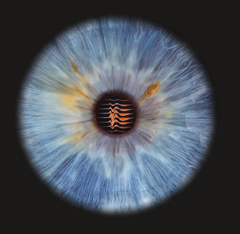
Picture: The picture is credited to Charité | Jens Kremkow & Fotostudio Farbtonwerk I Bernhardt Hyperlink
Authentic Analysis: Open entry.
“Excessive-density electrode recordings reveal sturdy and particular connections between retinal ganglion cells and midbrain neurons” by Jens Kremkow et al. Nature Communications
Summary
Excessive-density electrode recordings reveal sturdy and particular connections between retinal ganglion cells and midbrain neurons
The superior colliculus is a midbrain construction that performs vital roles in visually guided behaviors in mammals. Neurons within the superior colliculus obtain inputs from retinal ganglion cells however how these inputs are built-in in vivo is unknown.
Right here, we found that high-density electrodes concurrently seize the exercise of retinal axons and their postsynaptic goal neurons within the superior colliculus, in vivo.
We present that retinal ganglion cell axons within the mouse present a single cell exact illustration of the retina as enter to superior colliculus.
This isomorphic mapping builds the scaffold for exact retinotopic wiring and functionally particular connection power. Our strategies are broadly relevant, which we show by recording retinal inputs within the optic tectum in zebra finches.
We discover frequent wiring guidelines in mice and zebra finches that present a exact illustration of the visible world encoded in retinal ganglion cells connections to neurons in retinorecipient areas.

