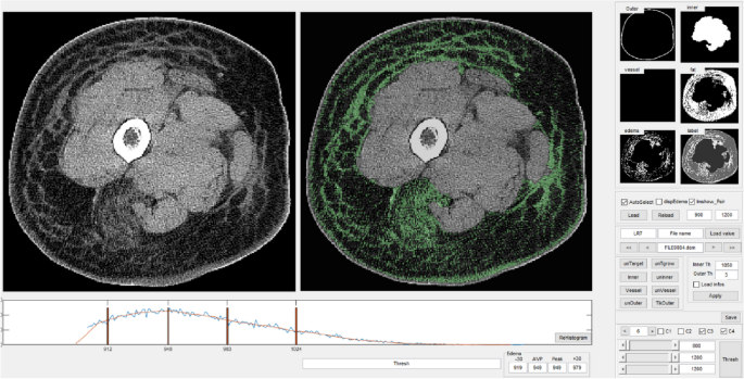
To indicate the validity of the DL-based algorithm for the automated quantification of lymphedema-induced fibrosis in CT pictures, a cross-sectional, observational comparability trial was performed in persistent lymphedema sufferers (ISL phases II to IIIa). The key findings are as follows: (1) the accuracy and MeanBFScore of the SegNet-based algorithm had been 0.776 and 0.868, respectively, displaying comparable energy to earlier trials making use of a CNN to different ailments; (2) the bulk (73.7%) of the 19 subindices of the 4 indices was considerably correlated with the BEI (partial correlation coefficient: 0.420–0.875), and the minority (13.2%) was considerably associated with the SCDR (partial correlation coefficient: 0.406–0.460); and (3) the imply worth of Index 2 (left( {frac{{P_{Fibrosis; in; Affected} – P_{Fibrosis; in; Unaffected} }}{{P_{Limb; in; Unaffected;} }}} proper)) within the distal a part of the limb, the place the subtraction methodology was used for standardization, introduced the strongest correlation with the BEI.
Ensembles of classifiers demand predefinition, resembling characteristic extraction and area of curiosity (ROI) definition36; thus, low accuracy and incomprehensive outcomes often happen. At present, numerous DL-based algorithms have been developed worldwide due to their strengths (i.e., picture segmentation and automatic characteristic era capabilities)37. CNNs, profitable DL algorithms primarily based on a multilayer hierarchical community, present excessive analytical efficiency when utilized to the medical pictures of sufferers with numerous sorts of ailments, resembling pulmonary tuberculosis38, breast most cancers39, mind tumors40, and hepatic ailments41. Nonetheless, just one examine utilized a CNN to the quantification of lymphedema-induced fibrosis. As a result of dependable, repeatable, and extremely correct strategies for the early detection of fibrosis (one of the vital persistent issues in illness administration) would have vital medical significance for the administration of lymphedema sufferers, there’s nice demand for the institution of such strategies. Constructive trials of DL-based picture classifiers, resembling AlexNet, VGGNet, U-Web, GoogLeNet, SegNet and ImageNet, would possibly result in success of this demand. Nonetheless, by way of semantic segmentation, U-Web and SegNet are the preferred42. SegNet, an computerized picture encoder-decoder, was developed for picture classification/picture segmentation in 201743. It was chosen right here as a result of it has less complicated layer construction than the U-Web in addition to this investigation required pixel-wise semantic classification within the CT picture. The authors have plan to implement the proposed segmentation utilizing U-Web and evaluate one another.
Previous to dialogue of SegNet software for CT-based fibrosis correlation with medical parameters, comparability of the present semiautomatic baseline methodology on the factor compositions’ segmentation would possibly higher justify the effectiveness of the proposed workflow. In our earlier trial with the identical undertaking, by which the present semiautomatic fibrosis segmentation methodology was used with the identical GUI (MATLAB [MathWorks, USA]), 3 varieties of CT fibrosis index formulated to judge their consultant functionality confirmed vital correlation with ISL substages (r of 0.68–0.79, p < 0.01), BEI ratio (r of − 0.46, p < 0.05), and proximal SCDR (r of 0.45, p < 0.05) and sensitivity of 0.78 and specificity of 0.60 in lymphatic system dysfunction detection21. By way of CT-based lymphedema fibrosis segmentation, just one report launched an identical methodology by which semiautomatic segmentation was performed by adjusting HU and adopted by a convex hull algorithm. Though correlative quantification of fibrosis with a third-dimensional quantity perometry outcomes failed, lateralization of the fibrosis areas was vital within the more-affected limb22. Equally, Edmunds et al. performed CT-based lean muscle space segmentation utilizing semiautomatic calculation of the variety of voxels with HU worth increased than that of fats. This calculation was adopted by smoothing and binning by a non-parametric becoming algorithm for quantification of muscle degeneration. They reported excessive correlation coefficients (r of 0.99, p < 0.005) with leg power, timed up-and go take a look at, gait velocity44.
Within the present trial, by which the picture knowledge had been divided into coaching (65%; 1252 pictures), validation (15%; 290 pictures), and testing (20%; 378 pictures) datasets and enter into SegNet, the accuracy, IoU, and MeanBFScore for fibrosis had been 0.776, 0.584, and 0.868, respectively. Regardless that the fibrosis accuracy (0.776) is decrease than different labels accuracy, it’s comparably increased than its random likelihood (1/5). It was suspected that similarity between the fibrosis and the fats in form has made it tougher.
Utilizing the identical CNN software, the accuracy of the SegNet-based segmentation mannequin for stroke lesion detection ranged from 85 to 87% within the mind MRI pictures of stroke sufferers45, by which the dataset contained 420 T1-weighted MRI scans divided into 2 units for coaching (70%; 294 pictures) and testing (30%; 126 pictures). In one other examine of 375 sufferers with 517 focal liver lesions (410 focal liver lesion pictures for coaching and 107 focal liver lesion pictures for testing), the mannequin confirmed increased accuracy within the detection of hepatocellular carcinoma and benign noninflammatory focal lesions (0.916 and 0.860, respectively) than that within the present trial46. Nonetheless, a special kind of CNN, particularly, a multiphase convolutional dense community (MP-CDN), was used. In one other examine utilizing SegNet-based chest X-ray picture segmentation (1674 pictures for coaching and 199 pictures for testing), the common accuracy for the detection of a lung nodule and the overlap rating had been 98.31% and 94.40%, respectively42. A comparability trial of a generative adversarial community (GAN) mannequin with a number of CNNs (12,150 belly CT pictures for coaching and eight,800 CT pictures for testing) confirmed that the SegNet IoU for hepatocellular carcinoma was 78.57, which is increased than the that within the present trial (0.584)47. All the aforementioned research, by which stroke lesions, liver lots, or lung nodules had been of curiosity, achieved a better accuracy and IoU than the present trial, by which fibrotic tissues had been of curiosity. This means that the DL-based quantification of lymphedema-induced fibrosis is a better problem. In comparison with the simple demarcation of oval- or semioval-shaped areas within the abovementioned trials, the troublesome demarcation of amorphous fibrotic tissues can clarify the decrease accuracy and IoU. Furthermore, the variations in demographic elements, resembling medical ailments, intercourse, and age, and the comparatively small pattern dimension of the present trial ought to be thought of.
In the meantime, hybrid DL fashions have proven very profitable leads to diagnostic accuracy because the early 2020s; in a CNN-based trial of Alzheimer’s illness mind MRI (2560 sections), options had been extracted by hybridization of Darknet53, InceptionV3, and Resent101 fashions and concatenated. Then, they had been optimized by mPMR and categorised by assist vector machine (SVM) and k-nearest neighbors (KNN) and confirmed very excessive accuracy of Alzheimer’s illness grades (none, very delicate, delicate, reasonable) starting from 94.4 to 99.1%33. In a CNN-based trial of cystourethrography on pediatric vesicoureteral reflux (1228 pictures), options had been extracted by hybridization of Googlenet, MobilenetV2, and Densenet201 fashions and concatenated. They then had been optimized by mPMR and categorised by SVM and KNN and confirmed very excessive accuracy of vesicoureteral reflux grades (regular to V) starting from 95.5 to 96.9%32. Relating to the comparatively low accuracy within the present trial, the latest hybrid DL fashions could be a possible answer to extend the accuracy.
Along with the aforementioned restricted accuracy, it’s unsure that the authors measured pure fibrosis. As proven in Figs. 3 and 4, what the authors referred to as “fibrosis” is areas with non-fat attenuation in subcutaneous fats layers. Such areas would possibly embody fibrosis but additionally can embody reasonably sized vessels and lymphedema itself. The authors are conscious of the inclusion of vessels and acknowledge that blood and surrounding vessels within the donut-shaped area had been labeled fibrosis primarily based on the idea that the full variety of blood vessel pixels (the full space occupied by blood vessels) was almost the identical within the affected and unaffected limbs.
Essentially the most vital downside is that the authors can’t discriminate between lymphedema and fibrosis. Fibrosis includes macroscopic change that may be visualized on histo-pathology10. As proven in earlier stories14,16,17, fibrosis may be seen on conventional-resolution CT at a 512 × 512 pixel matrix dimension and a component dimension of 0.5 mm (0.25 mm in super-high decision)48. Thus, fibrosis is measurable by present DL-based segmentation. In the meantime, exudate fluid in pure lymphedema with out fibrosis can induce reticulation in CT scanning49. So the reticulation within the subcutaneous fats compartment often comprises each water and fibrosis. HU worth for suprafascial lymphedema is understood to vary from − 120 HU to − 100 HU50. Nonetheless, the HU was measured a number of occasions at selective areas inside subcutaneous fats compartments with no discrimination of lymphedema from the fats in such stories. Moreover, fats infiltration, an inevitable complication in lymphedema10, can’t be helped however included as effectively in such assessments. Because the outcomes, each background fats and fats infiltration trigger shifting of the chosen ROI worth down towards that of fats (− 70 HU to − 190 HU)34, much like Edmunds et al.’s report44 and Aubrey et al.’s report51. Nonetheless, if this affection is abolished with the help of the tactic within the present examine, worth of the chosen ROI will return to the unique worth of sentimental tissue (30 HU to 60 HU)34,51. As well as, muscle tissue typically present CT worth of 30 HU to 60 HU in wholesome younger inhabitants51. Nonetheless, low attenuation of muscle tissue (− 29 HU to 29 HU) can occur in case of muscle-affecting focal/systemic ailments: for example, denervated muscle tissue and muscle tissue within the aged. In such circumstances, fats infiltration is probably the most acceptable issue as an underlying trigger51. Just like that, it’s well-known that fats infiltration happens contained in the affected muscle tissue because the outcomes of lymphedema-induced inflammatory cascade. Contemplating the aforementioned two findings, authors chosen pixels of HU from − 34 to 26 to take away the fat-infiltrated, low attenuated muscle tissue and interstitial water (HU: 0) contained in the lymphedema, after which reticulation pixels had been differentiated, through k-means clustering methodology, into both of the subcutaneous fats and the fibrosis within the present examine.
Nonetheless, lymphedema is outlined as accumulation of tissue fluid within the extracellular, interstitial house and may be noticed solely at a microscopic stage. CT, together with super-high-resolution CT, can’t but visualize a microscopic scale. Following the sample of improved MRI decision to 7.0 T and 11.74 T52,53, the authors anticipate advances in CT expertise to permit visualization of lymphedema.
In distinction to fibrosis segmentation efficiency, the correlation of the calculated indices with the medical gold normal measurements confirmed extra superior outcomes (Desk 3); earlier CT picture values confirmed a correlation coefficient of 0.46 with the 1-kHz-based BEI ratio and of 0.45 with the SCDR in a manually predefined segmentation examine of lymphedema-induced fibrosis21. The 0.45 correlation coefficient with the SCDR is much like that within the present trial (0.448 for the proximal SCDR and 0.406 for the distal SCDR). Nonetheless, the correlation coefficient (0.875) with the 1-kHz-based BEI ratio within the present trial is way increased than that (0.46) within the former trial. Contemplating that the aim of the present trial is to develop a DL-based algorithm as an alternative choice to conventional filter-based algorithms demanding guide predefinition and have extraction, this development suggests the prevalence of CNN over conventional strategies in computerized lymphedema-induced fibrosis recognition.
In lymphedema, inflammatory fibrosis is first triggered on the cellular-molecular stage through elevated transcription of IL-6, IL-8, TNF-α, and TGF-β1. If lymphatic system dysfunction doesn’t spontaneously get better or is effectively compensated following this set off within the acute interval54,55,56, lymphoproliferative irritation will proceed to worsen over time. This may end in macroscopic fibrosis consisting of collagen and extracellular matrix deposition10,11. Lymphedema-induced fibrosis takes place internally between the pores and skin and muscle layers by way of substitute of regular anatomical parts by fibrotic tissues somewhat than outward enlargement of the limb quantity10,11. Furthermore, this reconstitution change happens dominantly in subcutaneous fats layers57 within the type of a spatiotemporal cascade22. Due to this fact, a easy measurement of the exterior circumference can’t exactly signify this type of inside fibrotic substitute. Moreover, adiposity may be affected by anatomical location and intercourse58; the bigger is the realm of the concerned subcutaneous fats compartment in a person affected person, the better may be the discrepancy between the inside change and exterior limb circumference. Taking the above findings into consideration, a decrease correlation of the 4 indices with the SCDR could be anticipated. Furthermore, the low-frequency direct present utilized in BEI evaluation is principally performed through interstitial fluid59,60; in different phrases, the impedance worth measured by BEI evaluation, which depends on the relative ratio of connective tissue, by which electrical present can’t propagate as effectively as within the interstitial house, could possibly be extra appropriate for representing inside fibrotic modifications than the exterior circumference61. Contemplating the above findings, the excessive correlation of the 4 indices with the BEI could be anticipated. Nonetheless, additional comparability research involving direct histological findings, resembling these of tissue biopsies, could be required for extra particular validation.
All of the subindices of Index 2 confirmed reasonable to robust vital correlations with the BEI (0.630–0.875). Nonetheless, solely half of the Index 1, 3, and 4 subindices confirmed reasonable vital correlations (0.420–0.701). This discrepancy was exceptional. As a result of the pixels categorised as fibrosis within the affected limb had been subtracted from the pixels categorised as fibrosis within the unaffected limb, some segmentation errors within the affected limb may need been eradicated, doubtlessly contributing to the stronger correlation. In image-based medical trials, as a result of frequent phenomena of anatomical variation amongst people62, use of the relative ratio somewhat than absolutely the ratio is the popular knowledge standardization approach. Due to this fact, Index 2 calculated utilizing the above standardization methodology would possibly show increased classification energy than Indices 1, 3, and 4, such that it will present the best relationship with the BEI, much like a calculation formulation utilized in one other picture segmentation-based AI lymphedema examine21.
Limitations
As an alternative of an intention-to-treat evaluation, a per-protocol evaluation was used within the present trial as a result of the protocol was violated in 3 sufferers. The impact dimension may be overvalued in per-protocol analyses63. Furthermore, if the pattern dimension is small, this phenomenon may be stronger. As a broadly reported DL-related situation, overfitting (good match on the coaching dataset however poor efficiency on a brand new take a look at dataset) ought to be thought of64. As a result of pictures from 30 sufferers (1920 CT pictures) had been utilized for the mannequin growth within the present trial, actions to keep away from overfitting, resembling normalized layer era, dropout layer insertion, and knowledge augmentation to compensate for knowledge variations46, needed to be performed. As a result of pictures may be influenced by CT scanning parameters or acquisition protocols at particular person medical facilities, utilizing the present mannequin with a single CT scanner at a single heart may result in knowledge and/or mannequin bias. One other limitation is that feminine sufferers made up the overwhelming majority of the examine inhabitants (just one affected person was male)65. Moreover, the examine adopted a cross-sectional somewhat than potential design. Though lymphedema-induced subfascial muscular fibrosis66 and subsequent muscular change44 have been reported, they weren’t included right here. Future research are wanted to assist the present findings in a bigger variety of topics.


