Results of inoculum measurement on AMP manufacturing
The hemocyte-free hemolymph was checked for antibacterial activityon LB agar plates seeded with P. aeruginosa (ATCC 25668). All of the hemolymph samples had been collected at 4 h, 16 h, and 24 h from totally different batches of contaminated and non-infected B. mori. In consequence, all of the management group samples didn’t exhibit any antibacterial exercise. Furthermore, hemolymph collected after 4 h or 16 h of an infection didn’t exhibit any zone of inhibition. In contrast, hemolymph collected after 24 h of an infection confirmed zone of inhibition suggesting it’s inoculum-dependent and time-dependet exercise, which can be correlated with AMPs manufacturing. Additionaly, we noticed that every one the three bacterial hundreds (0.375 × 10⁸ CFU mL−1 to 1.5 × 10⁸ CFU mL−1) triggered AMP manufacturing with totally different inhibition zones (Desk 1). It was noticed that hemolymph remoted from the silkworm contaminated with 1.5 × 10⁸ CFU mL−1 for twenty-four h exhibited the very best antibacterial exercise in opposition to the examined bacterium. Thus, this contaminated group was chosen for AMP purification and characterization.
Adjustments in physio-morpho-anatomical responses in B. mori throughout bacterial an infection
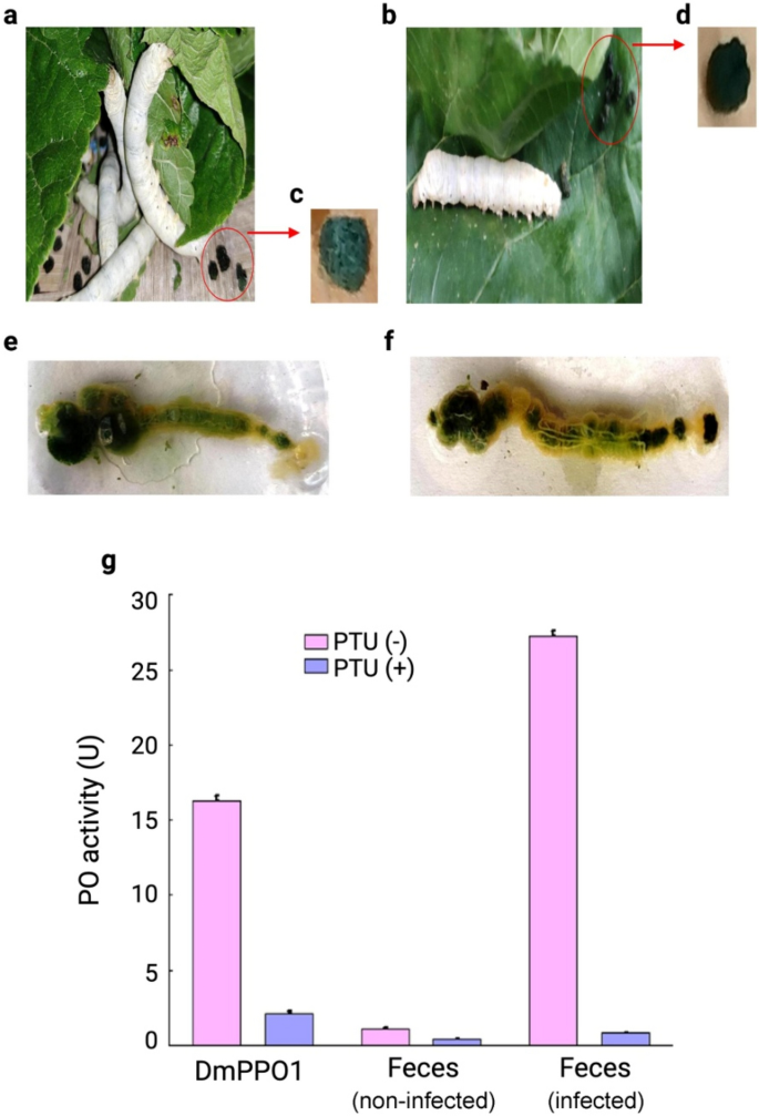
The current investigation revealed that non-infected group silkworm larvae consumed S1635 mulberry leaves excreted green-colored feces, whereas the contaminated group excreted black-colored feces (Fig. 2a–d). After dissecting the third day of fifth instar contaminated or non-infected larvae (24 h), we noticed that the alimentary canal content material of non-infected silkworm was inexperienced (Fig. 2e). In contrast, the alimentary canal content material of contaminated silkworm was discovered to be well-darkened in the direction of the cephalo-caudal finish (Fig. 2f). Melanization immune response is a principal component of insect immune protection in opposition to invading pathogens13,31,32. Following the microbial invasion, PPO is transformed into energetic phenoloxidase (PO), a course of mediated by serine proteases (SPs)33,34,35,36. PO participates in overseas pathogens elimination by Toll pathway activation34. PO is a crucial enzyme in B. mori that induces melanization cascades at wounds or across the invading pathogen37. Melanization is correlated with excessive PO exercise in hindgut content material and the darkening of feces. Right here, the enzymatic assay revealed that, when dopamine (phenoloxidase substrate) was incubated with D. melanogaster DmPPO1 within the absence of PTU (PPO inhibitor), excessive oxidative enzyme exercise by DmPPO1 is noticed (Fig. 2g). Nonetheless, within the presence of an inhibitor, DmPPO1 loses its exercise to a considerably low stage. Inexperienced-colored feces from non-infected silkworm larvae didn’t present any important enzymatic exercise, confirming the absence of PPO (Fig. 2g).
Fecal melanization in non-infected and contaminated B. mori larvae. (a) Non-infected larvae with continued feeding, (b) contaminated larvae that stoppted consuming, (c) green-colored feces of non-infected larvae, (d) black-colored feces of contaminated larvae, (e) dissection confirmed inexperienced intestine content material within the non-infected larval group, (f) black intestine content material within the contaminated larval group, and (g) polyphenol oxidase (PPO) exercise of non-infected and contaminated silkworm feces. The info had been expressed as imply ± customary deviation and the importance stage was set at p < 0.05.
Black-colored feces from contaminated larvae exhibited very excessive phenoloxidase exercise (p < 0.001) that, in flip, oxidized dopamine. This enzymatic exercise was considerably misplaced within the presence of PTU validate the presence of PPO within the intestine content material and moist feces of contaminated larvae, ensuing within the melanization and, in the end, blackening of feces. Turn into the inexperienced to black shade of feces can also be attributable to dehydration of hindgut content material, which can be attributed because of the polymerization of hydrophobic polyphenols into the feces. Together with the poisonous properties of oxidized phenol, the dehydration of intestine content material could be liable for lowering the pathogen’s load13. It’s identified that melanization in contaminated silkworm confirms the innate immune response attributable to bacterial invasion, which regularly prompts protein kinase cascade via Toll and IMD pathways to speed up AMP manufacturing (Fig. 1)1,3,38.
Our examine confirmed that silkworm larvae challenged with P. aeruginosa additionally induced modification in physiological processes, resulting in important morpho-anatomical adjustments. It was famous that contaminated silkworms instantly stopped feeding, which can be attributable to mechanical shock, whereas the non-infected silkworm confirmed regular foraging conduct (Supplementary Fig. S1a–b). It was additionally noticed that the contaminated larvae confirmed swelling within the head and thorax area (Supplementary Fig. S1c). On the second day of an infection, there have been adjustments within the integument shade from white to pale yellow (Supplementary Fig. S1d). On the third day of an infection, the integument was coffee-brown shade (Supplementary Fig. S1e) with minimal foraging conduct. Through the fourth and fifth days, the larval outer integument turned black, in the end ruptured, and died attributable to hemolymph leakage (Supplementary Fig. S1f).
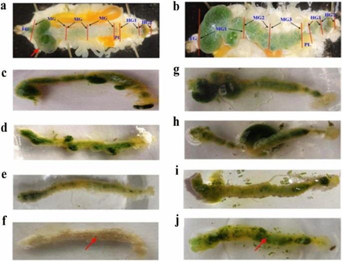
Our comparative anatomical examine revealed that, on the second day of an infection, there was an accumulation of meals content material within the foreguts (FG) and the primary a part of the midgut (MG1) with traces of melanized feces within the hindgut (HG). As a comparability, the non-infected silkworms had been additionally dissected (Fig. 3a,b). We additional recorded that the intestine content material regularly turned from inexperienced to white because of the modulation in foraging conduct and urge for food loss in contaminated silkworms from the primary to the fourth day of an infection (Fig. 3c–f). Management non-infected silkworms exhibited no intestine shade adjustments (Fig. 3g–j).
Anatomical comparability between contaminated and non-infected B. mori larvae. (a) Pink arrow displaying intestine content material accumulation within the contaminated larvae. (b) Management larvae confirmed inexperienced intestine content material all through the alimentary canal (FG, MG1, MG2, MG3, PL, HG1, and HG2). (c–f) The inexperienced shade of intestine content material within the alimentary canal decreased regularly and appeared full white in fourth day of contaminated larvae. (g–j) Management larvae confirmed no adjustments in intestine shade.
The urge for food loss triggered by bacterial an infection trigger important lower in each moist intestine weight and silk gland in comparison with the management group (Supplementary Fig. S2a–b). Our examine additionally revealed that because of the modulation in foraging conduct within the handled group, there’s a lower in silk manufacturing leading to white shade silk gland (Supplementary Fig. S2c–f). In contrast, the silk gland of the dissected non-infected group confirmed a gradual enhance within the depth of yellow shade, which might be attributable to regular feeding conduct accompanied by elevated silk manufacturing (Supplementary Fig. S2g–j).
As reported earlier, the pathogenic an infection causes important lower in larval physique weight, somatic index of silk gland tissue and weight of silk gland, which helps our findings39,40. A separate examine on PPO exercise in B. mori revealed that pathogenic an infection results in the immune induction through PPO activation cascade41. After pathogenic an infection, hindgut cells of silkworm larvae produces PO, triggering blackening and dehydration of each hind intestine content material and feces13. One other examine confirmed that knock-down and knock-out of PPO gene critically alters its immune capability and longevity in bugs, thus indicating the importance of insect PPO in immune safety and it’s correlation with AMPs manufacturing42.
UV–vis and FTIR examine of MGW extracted hemolymph
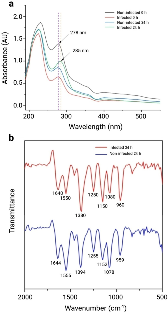
The protein spine includes a peptide chain with fragrant amino acids, together with tyrosine, tryptophan, and phenylalanine, which may soak up within the UV area due to the presence of π electron cloud. The absorbance of ultraviolet radiation at 280 nm signifies the presence of amino acids having fragrant rings and, typically, disulfide bonds to a small extent43. Furthermore, the peaks round 220 nm correspond to protein absorption. UV–vis spectra of non-infected hemolymph and contaminated hemolymph of B. mori after 0 h and 24 h, respectively, was recorded. The UV absorption peak at 278 nm was noticed for non-infected 0 h, contaminated 0 h, and non-infected 24 h. Nonetheless, a pointy change of λmax from 278 to 285 nm (bathochromic shift) was noticed for twenty-four h incubated contaminated silkworm hemolymph, which could be attributed to its chemical adjustments throughout bacterial an infection (Fig. 4a).
FTIR spectroscopy is a broadly used technique to research the construction of proteins and peptides both remoted or in a posh pattern (e.g., insect’s hemolymph). The FTIR spectra of non-infected and contaminated hemolymph from B. mori confirmed traits peak of amide bonds at totally different areas. The peaks at 1644 cm−1 and 1640 cm−1 symbolize the attribute peak of C = O stretching vibration of the amide I band of non-infected 24 h and contaminated 24 h hemolymph, respectively (Fig. 4b)44. Equally, the peaks at 1555 cm−1 and 1550 cm−1 symbolize the amide II band of non-infected 24 h and contaminated 24 h hemolymph, respectively (Fig. 4b). Moreover, two peaks at 1255 and 1250 cm−1 point out the presence of amide III absorptions, that are very weak in infrared spectra and come up from N–H bending44. A transparent shift of the three traits areas of doable peptide chain of non-infected 24 h and contaminated 24 h hemolymph is attributed to some chemical adjustments throughout the an infection. No peak was discovered at 1700–1750 cm−1, indicating the C-terminal amino acid in doable peptide chains shouldn’t be esterified. The height at 1550 cm−1 of contaminated 24 h strongly signifies the presence of α-helical peptides (with doable antibacterial properties) within the hemolymph. Lastly, the absorption at comparatively decrease wave numbers suggests the presence of water within the hemolymph45,46.
HPLC and Mass spectrometry
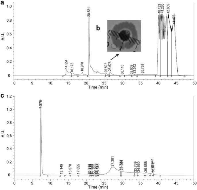
The MGW extracted hemolymph was purified via RP-HPLC for 50 min. The HPLC chromatogram (Fig. 5a) exhibits a number of peaks, the place every one was collected and examined for antimicrobial exercise in opposition to P. aeruginosa (ATCC 25,668). The fractions collected at 26.67 min (fraction 6) confirmed antimicrobial exercise (inhibition zone of 1.9 cm) in opposition to the examined bacterium (Fig. 5b) and, subsequently, was chosen for a second spherical of purification via RP-HPLC (Fig. 5c). The MIC worth of the remoted peptide was discovered to be 2 ppm in opposition to P. aeruginosa (ATCC 25,668). This MIC is in settlement with the outcomes obtained from earlier research, the place the antibacterial of quite a few AMPs, together with Q53, BmCec B, PA13, E53 BmCec B, Melimine and Mel4 had been decided in opposition to P. aeruginosa at 2.2 ppm, 3.91 ppm, 4 ppm, 66 ppm and 106.5 ppm, respectively47,48,49.
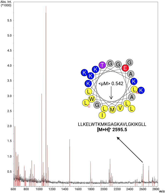
The fractions had been collected from 25 to 29 min and analyzed through Matrix-Assisted Laser Desorption Ionization (MALDI), aiming at characterizing the purified peptide. Among the many ions submitted to MASCOT evaluation, a peptide with molecular mass MH + 2595.61 Da was obtained (Fig. 6). This ion corresponds to a 24-amino acid residues peptide (LLKELWTKMKGAGKAVLGKIKGLL), as revealed via MALDI-TOF adopted by MASCOT evaluation. Right here, this peptide was initially named Bm-Frac6 (B. mori fraction 6) (Supplementary Desk S1). This peptide sequence introduced a whole match with an AMP deposited in APD, ponericin-L1 (APD ID: AP00383). This ponericin-like peptide was firstly remoted from ant venom and, attributable to its utterly id with Bm-Frac6 from this examine, we renamed our lead peptide candidate Bm-ponericin-L1 (B. mori ponericin-L1).
Characterization of Bm-ponericin-L1through MALDI-TOF. An ion of monoisotopic mass [M + H]+ of 2595.5 m/z is represented, similar to the peptide sequence LLKELWTKMKGAGKAVLGKIKGLL, beforehand described as ponericin L1 (an AMP remoted from ant venom). The helical wheel diagram can also be proven. < μM > = hydrophobic second.
Purified peptide in silico characterization
The peptide sequence described above was submitted to a set of computational analyzes. The Bm-ponericin-L1 peptide consists of a linear, cationic (internet cost + 5) peptide, with a hydrophobic charge of 45.8% and a hydrophobic second of 0.542 based mostly on the Eisenberg scale, thus indicating amphipathicity. Other than characterizing the physicochemical properties for Bm-ponericin-L1, we additionally retrived all of the 16 ponericin-like peptides deposited in APD and submitted their sequences to those self same calculations, as present in Supplementary Desk S2. Basically, the ponericin-like peptides current a net-positive cost starting from 2 to 7, total hydrophobicity from 22% to 60.8% (the latter is on the restrict worth acceptable for potential AMPs that don’t compromise the viability of wholesome mammalian cell traces), and hydrophobic second various from 0.228 to 0.587 (the upper the < μM > the larger the amphipathicity) (Supplementary Desk S2). When it comes to antibacterial properties, SVM, RF, ANN and DA algorithms from CAMPR3 predicted the Bm-ponericin-L1 peptide as a possible AMP (total chance = 99.4%). These predictions had been bolstered by the DBAASP prediction instrument, in addition to STM (prediction rating = 88.1%). Our computational information is additional strengthened by earlier experiences concerning the antimicrobial potential of the ant venom-derived ponericin-L150. Moreover, those self same algorithms had been utilized to the opposite ponericin-like sequences from APD, indicating that solely ponericin-G4 and ponericin-W6 didn’t attend the standards of ANN and DBAASP for antimicrobial exercise, respectively. Nonetheless, it’s price noting that every one the ponericin-like peptides had been predicted as AMPs by many of the prediction algorithms used, apart from ponericin-G4, which introduced the bottom possibilities and scores (Supplemantery Desk S3).
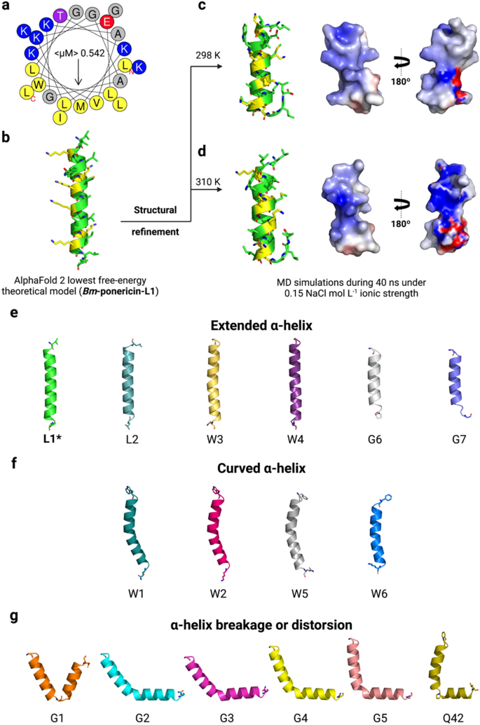
Contemplating the potential of Bm-ponericin-L1 as an AMP, additional computational research had been carried out to foretell its atomic coordinates, producing theoretical three-dimensional fashions. The molecular modeling was carried out utilizing the AlphaFold2 server, leading to a well-defined α-helical strcuture for Bm-ponericin-L1 (Fig. 7a,b). When it comes to construction statistics, the bottom free-energy mannequin forBm-ponericin-L1 presents all its amino acid residues in probably the most beneficial areas within the Ramachandran Plot (Supplementary Desk S4). Furthermore, the general common of the dihedral angles, together with the spine’s covalent forces resulted in a G-factor = 0.26 (> − 0.5 for dependable buildings) (Supplementary Desk S4). As for the physicochemical and antimicrobial prediction calculations, we prolonged our modeling evaluation to the opposite ponericin-like sequences retrieved from APD. The identical modeling strategies had been used and the theoretical fashions submitted to stereochemistry and fold high quality validations (Supplementary Desk S4). Amongst them, solely ponericin-G6 and -G7 confirmed lower than 90% of their amino acid residues within the Ramachandran Plot (Supplementary Desk S4). Apparently, nonetheless, when analyzing their secondary buildings we noticed three totally different structural profiles, together with a set of peptides (together with Bm-ponericin-L1) with prolonged α-helix (Fig. 7e), peptides with curved α-helix (Fig. 7f) and peptides presenting α-helix breakage or distorsions triggered by flexibility-inducer residues (e.g., glycine and proline) (Fig. 7g). To the perfect of our data, that is the primary report of the three-dimensional buildings of all ponericin-like peptides and their distribution into three structural scaffold “households”.
In silico structural characterization of Bm-ponericin-L1. (a) The helical wheel diagram for Bm-ponericin-L1is proven, together with its lowest free-energy three-dimensional theoretical mannequin. (b) The structural refinement was carried out at 298 (c) and 310 Okay (d), and 0.15 NaCl mol L−1 ionic power via MD simulations (40 ns). A complete of 16 ponericin-like peptides had been retrieved from APD and submitted to molecular modeling. These peptides had been divided into three primary lessons of construction profiles, together with (e) prolonged α-helix, (f) curved α-helix and (g) α-helix breakage or distorsions.
To enhance the accuracy of our in silico research, construction refinement simulations had been carried out for Bm-ponericin-L1 beneath two temperatures, 298 and 310 Okay, thus mimicking the totally different situations utilized in our in vitro experiments (Fig. 7c,d). The refinements had been accomplished utilizing MD simulations throughout 40 ns. As noticed in Fig. 7, the refined buildings are related and have a tendency to unfold on the C-terminal area because of the presence of a glycine residue at place 22. In consequence, the refined fashions for Bm-ponericin-L1, at each temperatures, current 16.6% much less α-helical content material than the non-refined construction. Furthermore, the basis imply sq. deviation (RMSD) between the Bm-ponericin-L1 refined buildings is 0.863 Å. When this identical evaluation is finished for the Bm-ponericin-L1 non-refined construction and its refined counterparts, a 1.4 Å total RMSD is noticed. In abstract, our computational research point out that Bm-ponericin-L1 presents a well-defined α-helical structural profile with a versatile C-terminus when analysed beneath 0.15 mol L−1 NaCl ionic power and 298 and 310 Okay.
Antibacterial and antibiofilm exercise
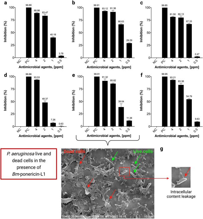
The antibacterial exercise of Bm-ponericin-L1was decided in opposition to ESKAPE pathogens and revealed a dose-dependent relationship. The outcomes confirmed that Bm-ponericin-L1 can inhibit the expansion of ESKAPE pathogens when in comparison with the bacterial progress within the detrimental management. The MIC (IC50) of Bm-ponericin-L1 ranged from 0.5 to 2 ppm for E. faecium, S. aureus, Okay. pneumoniae, P. aeruginosa, and E. agglomerans strains (Fig. 8a–c, e, and f); whereas, > 2 ppm was the MIC (IC50) for A. baumannii (Fig. 8d). Our examine additional revealed that there isn’t a strain-dependent variation within the antimicrobial exercise of Bm-ponericin-L1 when examined in opposition to P. aeruginosa (ATCC 10145) and P. aeruginosa (ATCC 25,668). SEM evaluation was carried out to guage the bactericidal results of Bm-ponericin-L1 on P.aeruginosa floor. Determine 8g exhibits the intact morphology with rod-shaped surfaces, as properly ascell membrane lysis brought on by Bm-ponericin-L1 remedy. The consequence signifies that Bm-ponericin-L1 presumably impacts intracellular metabolic reactions, inflicting induction in cytoplasmic injury or leakage (as highlighted). An analogous discovering was reported earlier for Gram-positive and Gram-negative micro organism handled with an defensin-like AMP derived from oyster51.
Impact of various focus of Bm-ponericin-L1 on ESKAPE pathogens. (a) E. faecium (ATCC 35667), (b) S. aureus (ATCC 6538), (c) Okay. pneumoniae (ATCC 70063), (d) A. baumannii (ATCC 17978), (e) P. aeruginosa (ATCC 10145), and (f) E. agglomerans (ATCC 27985). (g) SEM micrograph of intact/dwell micro organism (inexperienced arrows) and lysed/lifeless micro organism (purple arrows) within the presence of Bm-ponericin-L1. Adverse management (NC) and constructive management (PC). The info had been expressed as imply ± customary deviation and the importance stage was set at p < 0.05.
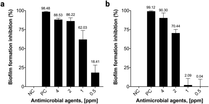
In vitro antibiofilm exercise of Bm-ponericin-L1 was measured at totally different concentrations in opposition to the biofilm-forming micro organism P.aeruginosa and Okay.pneumoniae. Our outcomes confirmed that Bm-ponericin-L1, at 4 ppm, inhibited biofilm formation by > 85% (P. aeruginosa) and > 90% (Okay. pneumoniae), after 24 h of incubation (Fig. 9a,b). Ourresult is comparable with an earlier examine that reported antibiofilm exercise of the insect-derived AMP (cecropin A) in opposition to uropathogenic Escherichia coli52.
Hemolytic and cytocompatibility assays
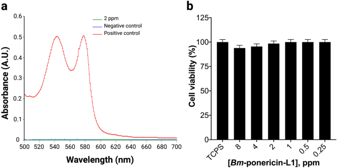
Hemolysis is a fundamental parameter to ascertain hemocompatibility of antimicrobials53. Our outcomes revealed that Bm-ponericin-L1 exhibited 0.2% hemolysis (Fig. 10a). It has been reported that antimicrobials with < 5% hemolysis had been considered hemocompatible54. Thus, the Bm-ponericin-L1 peptide, at 2 ppm, was discovered to be extra hemocompatiblethan different AMPs reported beforehand and which have proven hemolyisis in a dose-dependant method49. For this assay, diluted RBCs suspension blended with 0.8 mL PBS and 0.8 mL double distilled water had been used as detrimental and constructive management, respectively.
Hemocompatibility and cytocompatibility assay of the purified Bm-ponericin-L1 from contaminated B. mori larvae on erythrocytes (a) and first fibroblast cell traces (ATCC PCS-201-012) (b) respectively. The info had been expressed as imply ± customary deviation and the importance stage was set at p < 0.05.
To judge the results of the Bm-ponericin-L1 on the cell viability of major fibroblast cell traces (ATCC PCS-201-012), MTT assays had been performed with a variety of concentrations of the Bm-ponericin-L1 from 0.25 to eight ppm (Fig. 10b). The assay depends on the mitochondrial respiration system, which calculates the mobile vitality capability of a cell. MTT (yellow dye) is lowered by oxido-reductase and NADPH function electron donor producing a blue shade formazan. This transformation is just possible in viable cells; therefore, the amount of formazan is straight proportional to the variety of viable cells. In a separate examine, it was revealed that the insect-derived AMP cecropinXJ inhibited the expansion of Huh-7 cells in dose-dependent method, after 24 h of incubation. In that examine, cecropinXJ considerably inhibited the proliferation of Huh-7 cells with an inhibitory charge of ≤ 40% at 10 ppm55. By constrast, Bm-ponericin-L1 was non-toxic in the direction of major fibroblast cell traces (ATCC PCS-201-012) (Fig. 10b). Subsequently, Bm-ponericin-L1 particularly injury or kill bacterial cells, however is non-toxic in the direction of wholesome mammalian cells, as reported for different AMPs56.


